BALEEN IN MUSEUM COLLECTIONS: ITS SOURCES, USES, AND IDENTIFICATIONJULIE A. LAUFFENBURGER
2 BALEEN AND ITS SOURCES2.1 THE WHALE AND BALEENMammals of the Cetacea order, which includes whales, dolphins, and porpoises, can be divided into two suborders: Mysticeti and Odontoceti. The primary difference between Mysticeti and Odontoceti whales is their feeding mechanism. The Odontoceti are equipped with teeth to tear their food, and the Mysticeti have a special filtration system created by baleen. Baleen is a flexible horn-like material that grows in plates from the upper jaw of the Mysticeti (fig. 2). There are many species within this suborder, such as the humpback, finback, minke, sei, blue, and right whales. The more familiar sperm whale, immortalized by the writings of Herman Melville, is of the Odontoceti suborder.
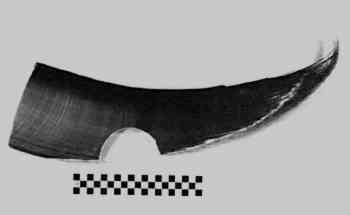
Baleen plates grow in parallel triangular sheets approximately 1 cm apart and can reach lengths of up to 13–14 ft (fig. 3). The plates grow continuously from the epidermal tissue of the gum and, like hair of other mammals, grow to maximum size sometime during adulthood. Seasonal variations in development result in visible growth ridges perpendicular to the length of each plate (fig. 4). When viewed from the exterior of the whale's mouth, the plates resemble rows of teeth on a comb. The lateral surfaces of baleen are flat, smooth, and resemble worked horn; the inner surfaces have hairlike fibrils. These fibrils are exposed only on the interior of the whale's mouth.
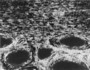
Sheets of baleen can vary in quantity and size depending on the age and species of the whale; bowhead whales are especially known for long baleen plates, which can exceed 700 in number. These plates also vary in quality, color, and relative fineness depending on the location of the plate in the whale's mouth and on the species. Baleen is generally cream, brown, gray, black, or white. Variation among plates can best be illustrated by comparing right whale baleen, which is extremely fine and gray in color, On the interior of the whale's mouth, the baleen fibrils act as a filter during feeding by forming a porous mat. Mysticeti whales feed primarily on small shrimp-like crustaceans called krill, which measure approximately 2 in long. The whale strains water filled with krill, other small crustaceans, squid, and fish by using its tongue to press against the “sieve” created by the frayed baleen fibers. Excess water and unwanted debris are expelled from the whale's mouth, while small krill and other edibles are trapped in the baleen mat. 2.2 STRUCTURE AND COMPOSITIONBaleen is a protein. Animal proteins are of two major categories: collagen and keratin. Collagen is an insoluble fibrous protein that is the chief constituent of connective tissues and bone. It is the primary component of leather and other skins. Keratin is a sulfur-containing fibrous protein Keratin is the major component of baleen. The complex keratin structure is held together by hydrogen bonds, polar linkages, and characteristically disulfide linkages of cystine (O'Connor 1987). Keratinaceous materials are divided into two groups: “soft” and “hard” keratins. Hard keratins have fibrils oriented along a major axis, giving them structure and strength, whereas soft keratins are randomly oriented proteins. Soft keratins exist in the continuously renewed outer layers of the epidermis of mammals, birds, and reptiles. Baleen and horn are both hard keratins and therefore have many of the same physical properties. Like horn, baleen can be softened in hot or boiling water. This process breaks down some of the disulfide bonds and allows the baleen to be pressed into a new conformation. When it is cooled under pressure, new bonds form, and the baleen retains its new shape. A plate of baleen is composed of a sandwich-like, three-part structure: a central section of tubules in a cementing keratin matrix, flanked by a horny covering. Figure 5 is a cross section taken from a plate of bowhead baleen. Inside the whale's mouth, the horny covering and cementing matrix are worn away by the abrasive action of the tongue, revealing the central tubules or hairlike structures that form the mat or sieve during filtration. These tubes are highly calcified, containing crystals of hydroxyapatite (a basic calcium phosphate) (O'Connor 1987), and they have central hollow cavities of varying sizes depending on the species. Calcification occurs in alternating rings around the outer edge of the tubes, an arrangement that strengthens the tubes without loss of flexibility (O'Connor 1987). Baleen is the most highly calcified of the keratin materials and as a result is slightly more brittle (Halstead 1974).
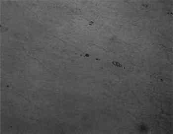
Artifacts made from complete plates of baleen can be easily identified because of the unique tripartite structure, but if only the
|
